Cross Section Of A Compact Bone / Bone Canaliculus Wikipedia / Spongy bone is used for more active functions of the bones, including blood cell production and ion exchange.
byAdmin-
0
Cross Section Of A Compact Bone / Bone Canaliculus Wikipedia / Spongy bone is used for more active functions of the bones, including blood cell production and ion exchange.. Use colored pencils to draw and label the following structures as they appear using the 40x objective, or by looking at an image from the internet. Fibrous astrocytes in white matter of the brain. Slides have to be made this way because the matrix of bone is too hard to Obtain a demineralized compact bone preparation (in cross section), preferably from the diaphysis of a long bone, and prepare to examine it microscopically. Smooth muscle fibers teased apart 2.
Compact bone, as opposed to spongy bone, is made of cylindrical units, called osteons, that are tightly formed together. They branch and anastomose and become continuous with volkmann's canals. Due to the strong nature of compact bone, compared to spongy bone, it is the preferred tissue for strength. It consists of two layers; Then, fill in the table below to describe each.
Anatomy Descriptive And Applied Anatomy Fig 5 Nucleated Bone Cells Osteoblasts And Their Processes Contained In The Bone Lacuna And Their Canaliculi Respectively From A Section Through The Vertebra Of An Adult from c8.alamy.com The first, outermost layer of membrane (besides periosteum) serves to. A cross section of a compact bone shows concentric circles called lamellae. A central tube called a haversian canal typically runs in the same path as the length of the bone. It is dense (because of calcified matrix) with tiny spaces known as lucanas. Obtain a demineralized compact bone preparation (in cross section), preferably from the diaphysis of a long bone, and prepare to examine it microscopically. Spongy bone is used for more active functions of the bones, including blood cell production and ion exchange. The compact bone is the main structure in the body for support, protection, and movement. That is, answer all cases of the question.
Skull bone is a flat bone.
Skull bone is a flat bone. As compact bone grows, osteons begin to fuse together. Label the haversian canal, osteocyte (mature bone cell) in lacuna, and canaliculi. The outlined area is a cross section of an osteon of compact bone. Moreover, it is a storehouse of calcium and hosphorus. This slide contained a cross section of a very small bone, and you are looking at the entire thickness of the shaft of the bone. A cross section of a compact bone shows concentric circles called lamellae. These are abundant and characteristic of compact bone. Fibrous astrocytes in white matter of the brain. Compact bone is made of concentric layers of osteocytes and bony matrix. In long bones, as you move from the outer cortical compact bone to the inner medullary cavity, the bone transitions to spongy bone. Compact bone is the outer layer and the spongy bone forms the inner layer. This is known as the periosteum.
The first, outermost layer of membrane (besides periosteum) serves to. Some, mostly older, compact bone is remodelled to form these haversian systems (or osteons).the osteocytes sit in their lacunae in concentric rings around a central haversian canal (which runs longitudinally).the osteocytes are arranged in concentric rings of bone matrix called lamellae (little plates), and their processes run in interconnecting canaliculi. Smooth muscle fibers teased apart 2. Within each lamella, collagen is mixed with inorganic minerals like magnesium, calcium and phosphorus and layered around a haversian canal. The osteon has blood vessels and bone cells, things vital for the survival of the bone.
Structure Of Compact Bone A Cross Sectional View Of Compact Bone Download Scientific Diagram from www.researchgate.net The compact bone is made up of osteon. Skeletal muscles showing _ fibers. The compact bone is the main structure in the body for support, protection, and movement. Because of its strength, the compact bone makes it possible for the bone to support weight. A cross section of a compact bone shows concentric circles called lamellae. Compact bone is very different from the other tissues you have seen. A cross section of a compact bone shows concentric circles called lamellae. Fibrous astrocytes in white matter of the brain.
Some, mostly older, compact bone is remodelled to form these haversian systems (or osteons).the osteocytes sit in their lacunae in concentric rings around a central haversian canal (which runs longitudinally).the osteocytes are arranged in concentric rings of bone matrix called lamellae (little plates), and their processes run in interconnecting canaliculi.
Compact bone ground cross section. ( ) each osteon has a central haversian canal , running parallel to long axis of bone. Compact bone is the outer layer and the spongy bone forms the inner layer. Then, fill in the table below to describe each. Skeletal muscles showing _ fibers. Due to the strong nature of compact bone, compared to spongy bone, it is the preferred tissue for strength. Also called cortical bone, the compact variety usually features a haversian system, or cylindrical unit within the structure. Skull bone is a flat bone. They fill the inner layer of most bones such as the vertebrae. In long bones, as you move from the outer cortical compact bone to. The large dark spots are passages for blood vessels and nerves. Before placing your slide on the microscope stage, remember to read the label, examine the slide with your eye and note any visible macroscopic features that might help your examination. The compact bone is the main structure in the body for support, protection, and movement.
As the names suggest compact bone looks compact and the spongy bone looks like skull bone is a flat bone. They branch and anastomose and become continuous with volkmann's canals. Compact bone is made of concentric layers of osteocytes and bony matrix. Related posts of cross section of a long bone bone test anatomy and physiology. Bone test anatomy and physiology 12 photos of the bone test anatomy and physiology anatomy and physiology bone lab test, anatomy and physiology bone markings test, anatomy and physiology bone practical test, anatomy and physiology bone tissue test, anatomy and physiology test on bone tissue, bone, anatomy and.
Bone Biopsy Open from content.ca.healthwise.net The first, outermost layer of membrane (besides periosteum) serves to. It consists of two layers; Skeletal muscles showing _ fibers. Observe that the matrix of the bone is deposited in concentric layers that are called lamellae (5). A cross section of a compact bone shows concentric circles called lamellae. Obtain a demineralized compact bone preparation (in cross section), preferably from the diaphysis of a long bone, and prepare to examine it microscopically. It is dense (because of calcified matrix) with tiny spaces known as lucanas. A cross section of a compact bone shows concentric circles called lamellae.
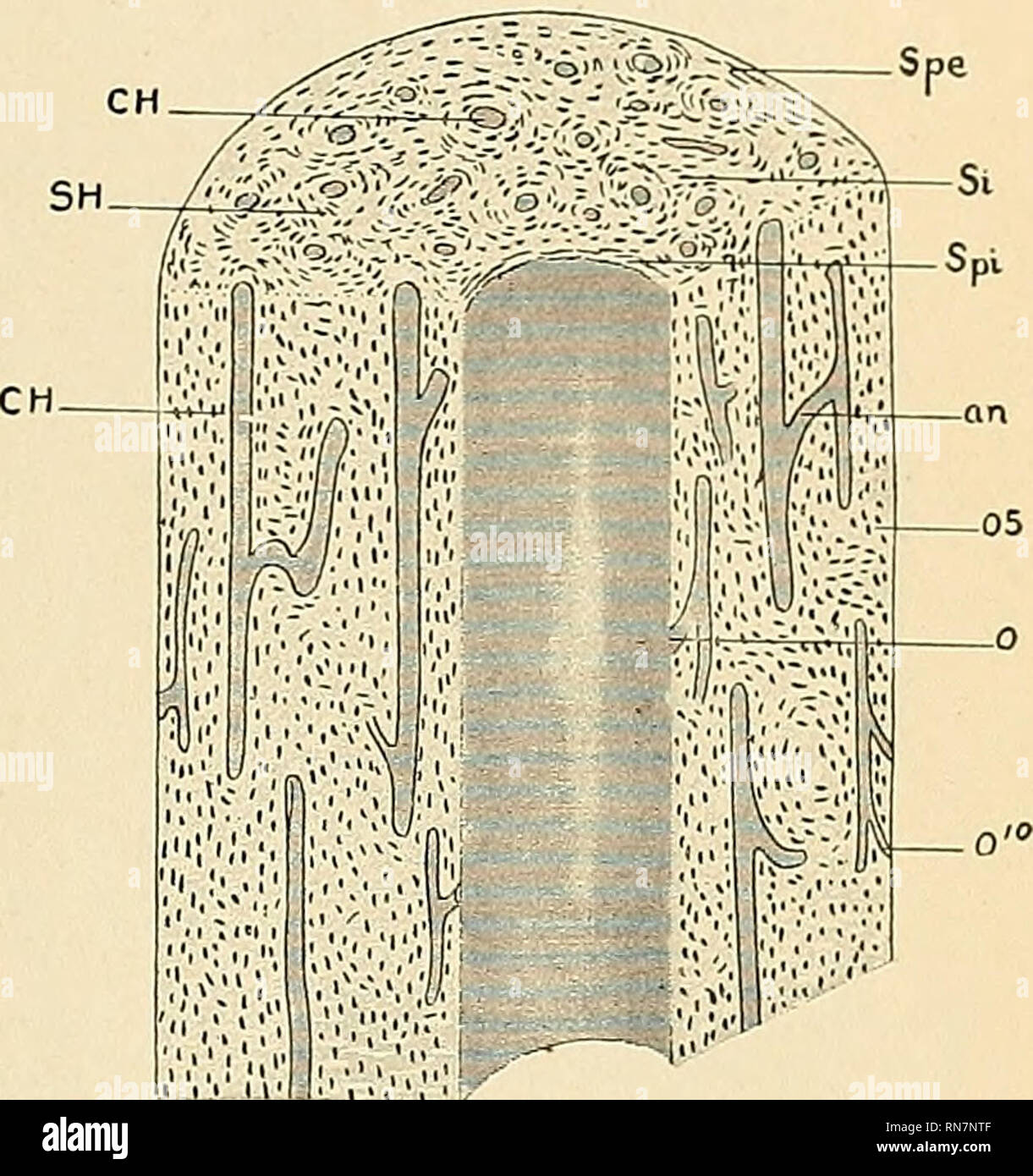
A diagrammatic view of a cross section of bone.
In the center of each osteon is the central canal, a space that houses blood vessels and nerves that supply bone. When compact bone is studied, it is found to be made up of concentric circles called lamellae. The outlined area is a cross section of an osteon of compact bone. A cross section of a compact bone shows concentric circles called lamellae. Obtain a demineralized compact bone preparation (in cross section), preferably from the diaphysis of a long bone, and prepare to examine it microscopically. Compact bone decalcified cross section. Moreover, it is a storehouse of calcium and hosphorus. These are abundant and characteristic of compact bone. Related posts of cross section of a long bone bone test anatomy and physiology. Use colored pencils to draw and label the following structures as they appear using the 40x objective, or by looking at an image from the internet. The remainder is cancellous bone, which has a spongelike appearance with numerous large spaces and is found in the. As compact bone grows, osteons begin to fuse together. Because of its strength, the compact bone makes it possible for the bone to support weight.
Compact bone is the outer layer and the spongy bone forms the inner layer cross section of a bone. Compact cross section human, ground bone, 162 x.